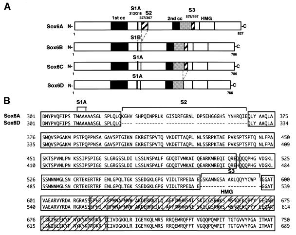
Figure 1.
Sequence comparison of SOX6 proteins. (A) Schematic comparison of SOX6 isoforms, SOX6A, SOX6B and SOX6C (28) together with the variant SOX6D. S1, S2 and S3 designate segments that differ between the isoforms. Two putative coiled-coil domains (1st cc and 2nd cc) and the HMG-box DNA binding domain common to all isoforms are indicated. (B) Comparison of the central region of SOX6A and SOX6D showing the divergent amino acid sequences. Identical residues are shaded.

