Figure 2.
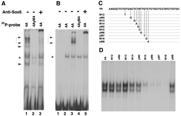
Binding and recognition sequence of SOX6 proteins in PS4A. (A) EMSA using in vitro transcribed and translated SOX6 proteins and 32P-labeled probes. Lanes 1 and 3, 4A; lane 2, mutated probe 4AyM4. The + indicates addition of SOX6 antiserum after complex formation. (B) EMSA using recombinant SOX6 protein alone (lane 1) or mixed with rabbit reticulocyte lysate (lanes 3–5). Rabbit reticulocyte lysate alone (lane 2). 32P-radiolabelled probes are indicated above the lanes. Positions of complexes and a non-specific band are indicated by arrowheads and an asterisk, respectively. The two smaller complexes [lane 1 in (A)] probably arise from initiation of translation at internal AUG codons. (C) Schematic depiction showing the sequences of wild-type and point mutated PS4A probes used to establish the SOX6 binding site. (D) EMSA using 32P-labelled PS4A probes shown in (C), mixed with purified GST-Sox6DB protein.

