Figure 6.
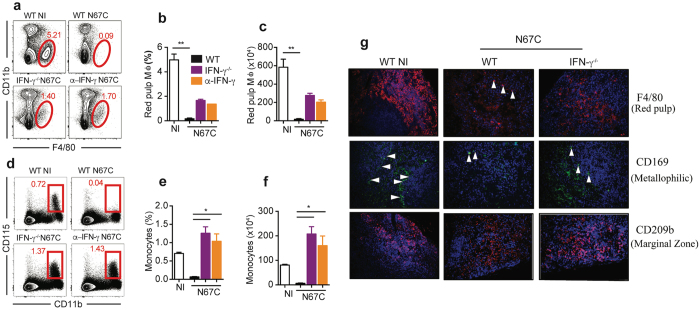
Retention of macrophages (Mɸ) subtypes and monocytes in IFN-γ−/− mice. (a) Representative flow cytometry plots of red pulp Mɸ (F4/80+ CD11b-) of WT, IFN-γ−/− and anti-IFN-γ mAb treated mice day 4 p.i. (b,c) Percentage and absolute number of red pulp Mɸ. (d) Representative flow cytometry plots of inflammatory monocytes (CD11b+ CD115+). (e,f) Percentage and absolute number of inflammatory monocytes. Data in (b,c and e,f) are mean ± SEM (3–5 mice) and are representative of two experiments; Kruskal-Wallis test, *p < 0.05, and ***p < 0.01. (g) Immunofluorescent staining of splenic Mɸ sub-populations using specific markers as described in40. The red pulp Mɸ was stained by F4/80 expression, marginal zone Mɸ by CD209b, and metallophilic Mɸ by CD169 (40X magnification). The arrowheads point to stained cells of different sub-populations of macrophages.

