
FIGURE 3.
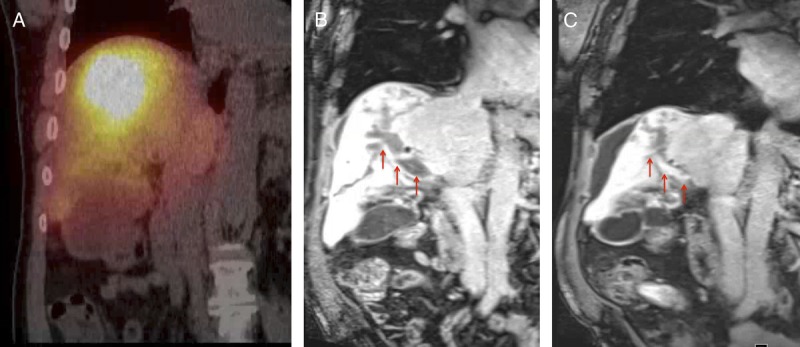
Case 2, Imaging findings. Postprocedural Bremsstrahlung fused SPECT-CT scan (A), demonstrates increased tracer activity within the expected location of the infiltrative lesion and within the right portal vein. On the correlative T1-weighted MRI image on portal venous phase on the preprocedural scan (B), the tumor thrombosis involves the right portal vein (arrow). In the correlative T1-weighted MRI image on portal venous phase on the 3-month postprocedural scan (c), note the partial recanalization of the portal vein (arrow).