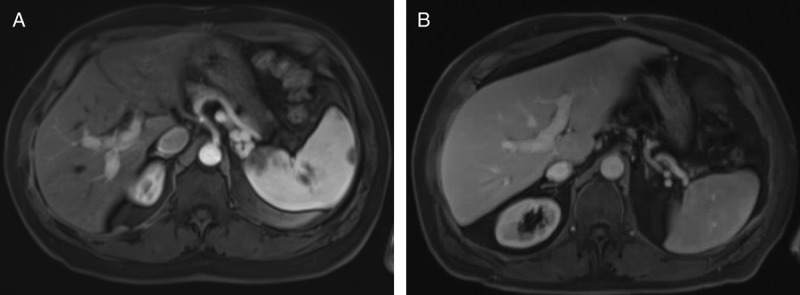
FIGURE 7.

Posttransplant imaging surveillance. Posttransplant axial contrast enhanced T1-weighted MRI image on delayed arterial phase. (A) Case 1 at 24 months, and (B) case 2 at 20 months, demonstrates no areas of arterial enhancement as well as patent portal veins.
