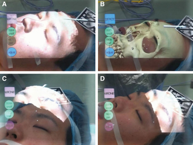
Fig. 8.

Actual shots through the AR device during the operation in a patient with an osteoma. A, The body surface is displayed. B, The facial bones including the osteoma are displayed. C, D, The body surface extracted only around the osteoma is displayed; it has been shifted from the corresponding surgical field. When the deformation is small, this method is easier to compare with the surgical field.
