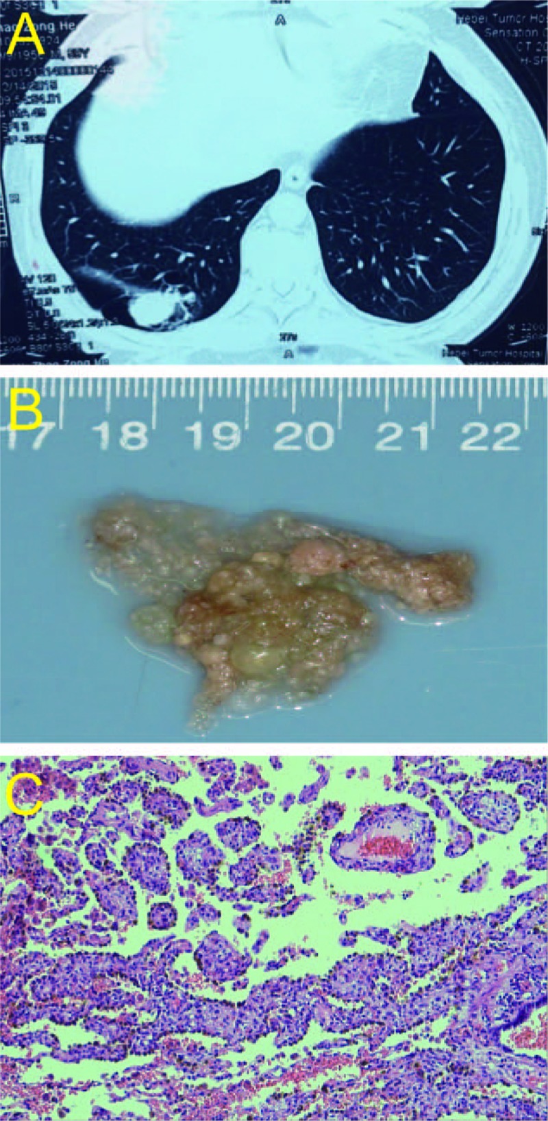
Figure 1.

Gross and microscopic features of PTL. (A) CT scan of PTL, the soft tissue mass of the right lower lung, measuring 3.8 cm × 2.33 cm. The edge of the tumor was clear, and the shallow lobe with multiple peripheral pulmonary bullae was found. (B) Gross pathology: well-circumscribed mass with papillary projections, resembling a placenta. (C) Photomicrograph with hematoxylin and eosin staining shows the cystic lesions of the lung containing multiple villous papillary structures similar to placental villi.
