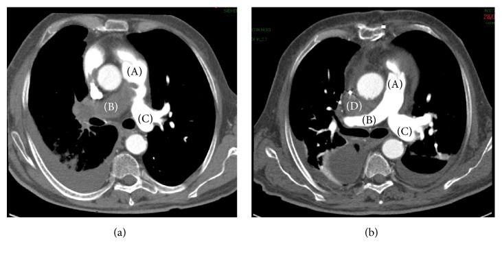
Figure 1.
CT scan of patient number 10. (a) Presurgical CT scan: right pulmonary artery completely occluded by soft tissue mass (B); proximal aspect of the mass in the pulmonary trunk (A); left pulmonary artery appears preserved (C). (b) Postsurgical CT scan (same level): the intra-arterial mass has been completely removed. Small mediastinal haematoma (D) between ascending aorta and right pulmonary artery.

