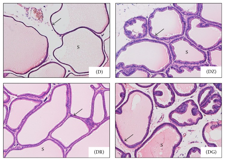
Figure 3.
Higher magnification of the preceding photomicrograph, ventral prostate in different groups; diabetic rats (D) and diabetic treated rats (DZ; DR; DG). Prostatic secretions (S) were darkly stained in diabetic treated groups when compared with diabetic ones; ductal walls (arrows) were clearly thicker in treated groups while, in diabetic group, they were thin (H&E ×100).

