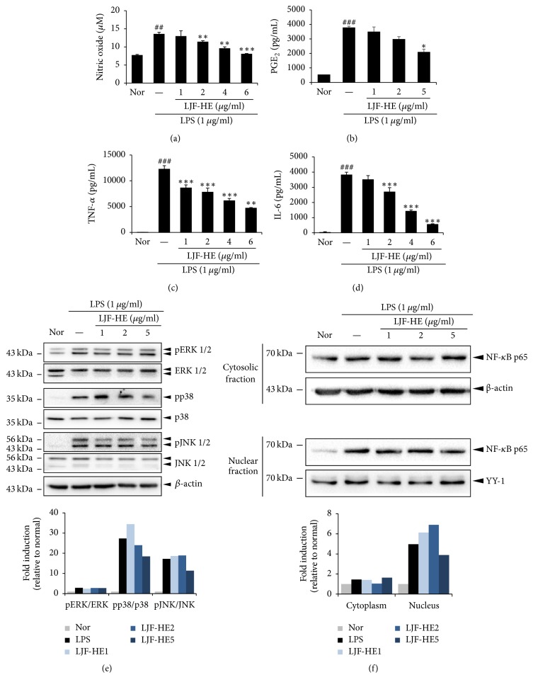
Figure 6.
Effects of LJF-HE on inflammation in LPS-stimulated RAW 264.7 cells. RAW 264.7 cells were pretreated with LJF-HE (1–6 μg/mL) for 1 h and then treated with LPS (1 μg/mL) for 0.5 h or 18 h. The levels of (a, b) NO and PGE2 in the cell culture supernatant were measured using NO and PGE2 detection kits. The levels of (c, d) TNF-α and IL-6 were measured by ELISA. (e) The phosphorylation levels of ERK1/2, JNK1/2, and p38 were analyzed by western blotting. The relative amount of each protein was determined using ImageJ software. (f) Nuclear, cytosolic, and total proteins were analyzed by western blotting using NF-κB p65 antibodies. β-Actin and YY-1 were used as internal controls for the cytosolic and nuclear proteins. The relative amount of each protein was determined using ImageJ software. ## indicates p < 0.01 and ### indicates p < 0.001 when compared to the untreated cells (Nor). ∗ indicates p < 0.05, ∗∗ indicates p < 0.01, and ∗∗∗ indicates p < 0.001 when compared to the LPS-treated cells.

