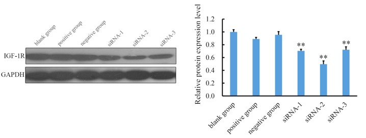
Figure 3.
Relative protein expression of IGF-1R in each group was detected by western blotting. Levels of IGF-1R in the three siRNA groups were lower than in the blank control group. Quantified data indicated that siRNA-2 exhibited the highest inhibition efficacy. All data are presented as the mean + standard deviation. **P<0.01 vs. blank control group. siRNA, small interfering RNA; IGF-1R, insulin-like growth factor receptor 1.

