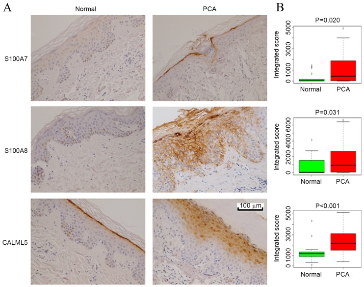
Figure 3.
IHC validation of selected differentially expressed proteins in PCA. (A) Representative images of IHC analysis. Skin samples from normal controls (n=17) and patients with PCA (n=29) were subjected to S100A7, S100A8 or CALML5 staining. (B) Box and whisker plots of integrated scores from the IHC staining of epidermal cells in the normal and PCA groups. S100A7/8, S100 calcium-binding protein A7/8; CALML5, calmodulin-like protein 5; IHC, immunohistochemistry; PCA, primary cutaneous amyloidosis.

