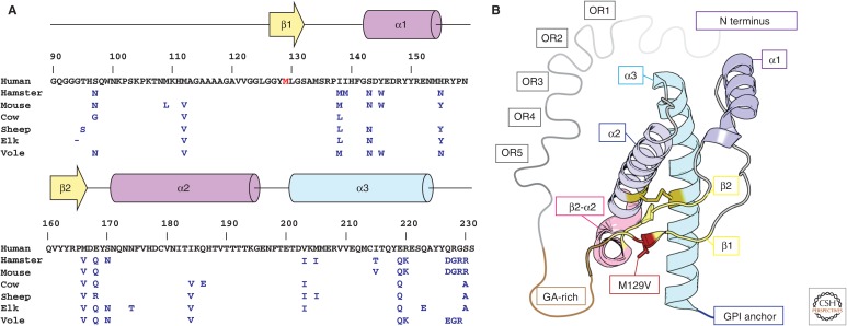
Figure 1.
Sequence and structure of human PrPC. (A) Primary sequence and secondary structure of human PrPC residues 90–230. Residues that differ between hamster, mouse, cow, sheep, elk, and bank vole sequences are indicated in blue, and those of the native human sequence are shown in black. The genetic variant M129V is shown in red. (B) Model of the crystal structure of the M129V variant of human PrPC. Secondary structure elements are labeled using a color scheme that follows A; these include five octapeptide repeats (OR1–5, gray), a glycine-alanine-rich stretch (GA-rich, brown), two β-strands (β1, β2, yellow), and three α-helices (α1–α3, magenta-blue), variant M129V (red), the β2–α2 turn (pink), and a C-terminal glycosylphosphatidylinositol (GPI) anchor (blue).

