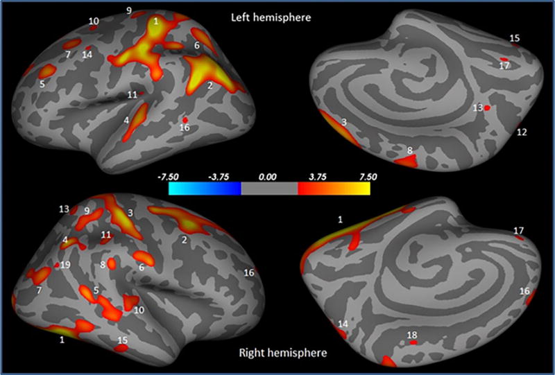
Fig. 1.

Brain regions showing significantly reduced cortical thickness in PD male compared to PD female overlaid onto inflated pial surface. These regions are postcentral (1), inferior parietal (2), superior frontal (3), superior temporal (4), rostral middle frontal (5), superior parietal (6), caudal middle frontal (7), precentral (8), superior frontal (9), superior frontal (10), supramarginal (11), superior parietal (12), precuneus (13), caudal middle frontal (14), fusiform (15), middle temporal (16), lingual (17) in the left hemisphere, and fusiform (1), caudal middle frontal (2), postcentral (3), inferior parietal (4), inferior parietal (5), postcentral (6), inferior parietal (7), supramarginal (8), superior parietal (9), superior temporal (10), supramarginal (11), superior frontal (12), superior parietal (13), superior parietal (14), inferior temporal (15), rostral middle frontal (16), medial orbitofrontal (17), paracentral (18), and inferior parietal (19) in the right hemisphere
