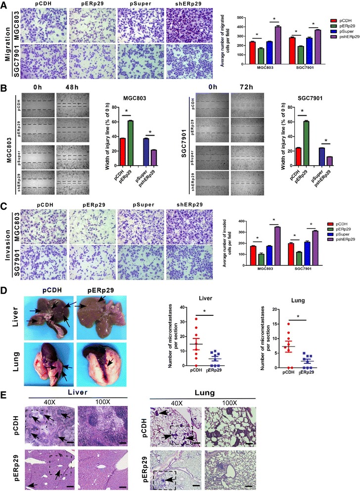
Fig. 3.

ERp29 regulated GC cell migration, invasion and metastatic potential. (a) Relative migration of the GC cells through an uncoated filter toward serum-containing medium in a Boyden chamber assay. (b) Relative motility as determined by the ability of the GC cells to close a wound made by creating a scratch through a lawn of confluent cells. (c) Relative invasion of cells through a layer of Matrigel coated on the filter of a Boyden chamber. (d) Quantification of liver and lung metastatic burden in mice 10 weeks after tail vein injection of the GC cells by counting the number of micrometastases per section. (e) Hematoxylin and eosin staining of fixed and paraffin-embedded tissues confirmed the presence of micrometastases in the liver (40 × magnification; scale bar: 50 μm) and lungs (100 × magnification; scale bar: 20 μm) of mice injected with the GC cells. *P < 0.05
