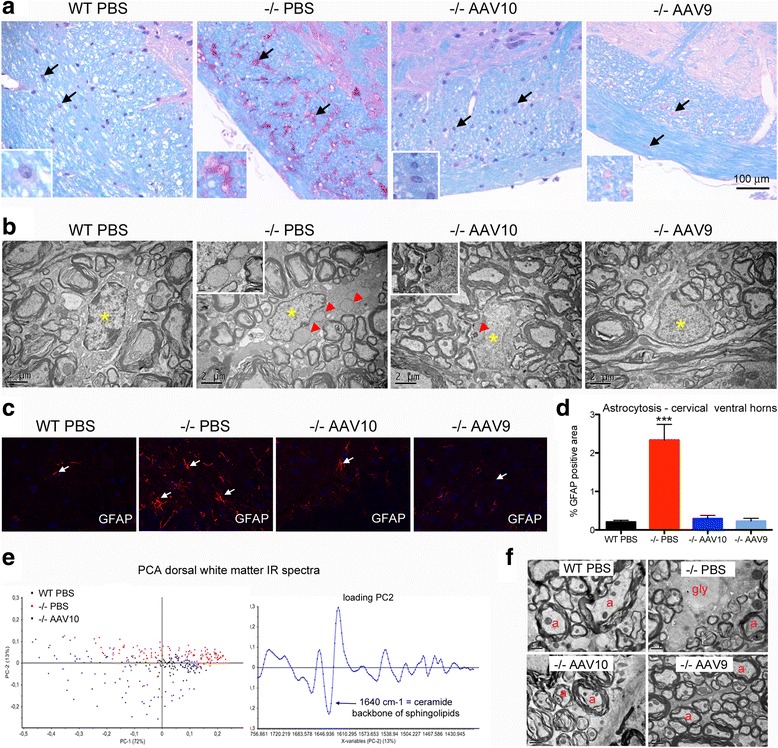
Fig. 4.

Glial cells and myelin are corrected at 12 months. Treatment groups were as described in Fig. 1. a Representative sections of brainstem peripheral white matter (spinal trigeminal tract), paraffin embedding, PAS-luxol fast blue stain. Arrows point to glial cells, astrocytes and oligodendrocytes. Insets show glial cells, presumably astrocytes, at a higher magnification (b) Representative ultrastructure of cervical spinal cord white matter oligodendrocytes, epon embedding, uranyl acetate contrast. The nuclei are indicated with yellow asterisks and the glycogenosomes with red arrowheads. c Representative immunofluorescence labeling of astrocytes using antibodies that recognize GFAP (glial fibrillary acidic protein) in cervical spinal cord frozen section (ventral horns). d Qualification of astrocytosis in cervical spinal cord ventral horns (one-way ANOVA with Newman-Keuls post hoc test; n = 4 per group: ***P < 0.001) (e) Principal component analysis (PCA) of the IR spectra collected by mapping spinal cord dorsal white matter in FTIR microspectroscopy using a synchrotron light source (n = 99, 149, and 94 spectra for WT, −/− PBS, and −/− AAV10 respectively) and corresponding loading plot. f Representative ultrastructure of cervical spinal cord dorsal white matter, epon embedding, uranyl acetate contrast. Myelinated axons (a), glycogen (gly) is seen in some demyelinated enlarged axons causing rarefaction of normal myelinated axon profiles in the mock-treated mice
