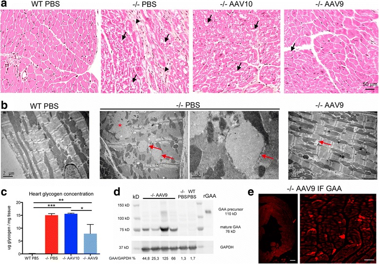
Fig. 7.

Cardiac muscle pathology improvement is correlated to the presence of GAA. a Representative cross sections of the left ventricular free wall, paraffin embedding, hemalun-eosin-saffron stain. The collagen appears orange on this trichrome stain. Arrows point to vacuoles in the cytoplasm of the fibers and arrowheads point to fibrosis. The cardiac muscle pathology is improved with AAV9 only. b Representative ultrastructure of the left ventricular free wall, epon embedding, uranyl-acetate contrast. Arrows point to glycogenosomes and the asterisk shows a “glycogen lake” that occurs after rupture of enlarged lysosomes. c Glycogen concentration measurement in heart extracts obtained from samples that were snap-frozen in liquid nitrogen rapidly after sacrifice (one-way ANOVA with Newman-Keuls post hoc test; n = 4 to 5 per group: **P < 0.01, ***P < 0.001) (d) Western-blot analysis of heart tissue extracts with a rat polyclonal anti-rGAA antibody. Glyceraldehyde-3-phosphate dehydrogenase (GAPDH) immunodetection is used as a protein loading control. e Representative immunofluorescence labeling of GAA in AAV9 treated mice using a rat polyclonal anti-rGAA antibody on a frozen heart section. Scale bars = 200 μm
