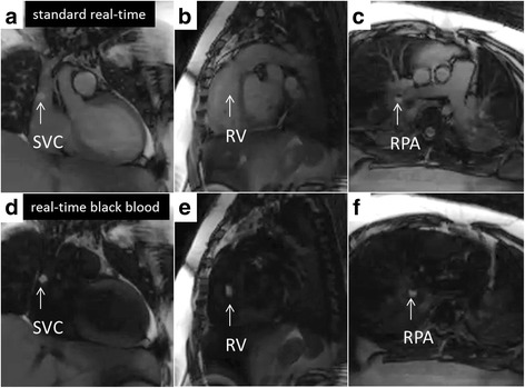
Fig. 3.

CMR right heart catheterization. Panels (a, b, and c) show real time CMR catheter navigation to superior vena cava (SVC), right ventricle (RV), and right pulmonary artery (RPA) respectively. Panels (d, e, and f) show the same respective imaging planes after flow-sensitive saturation preparation pulse to null blood pool. Gadolinium filled balloon (white arrow) is easily and often better (best represented in panel b versus panel e) visualized during this real-time black blood imaging
