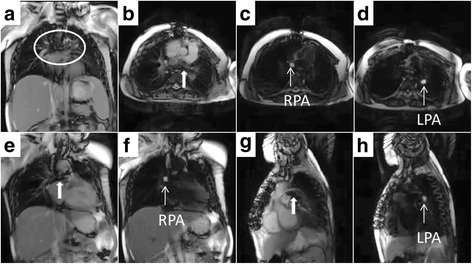
Fig. 4.

CMR cardiac catheterization in patient with pulmonary artery stent imaging artifact. Panel (a) shows imaging artifact (circled) from previously placed pulmonary artery stents; CMR right heart catheterization was successful. Panel (b) shows oblique axial imaging plane showing branch pulmonary arteries (thick white arrow = stent imaging artifact). Panel (e) and (g) show oblique coronal imaging planes for right and left pulmonary artery respectively. Panels c (RPA = right pulmonary artery), d (LPA = left pulmonary artery), f (RPA), h (LPA) show the same respective imaging planes after flow-sensitive saturation preparation pulse to null blood pool. Gadolinium filled balloon is easily visualized during this real-time black blood imaging
