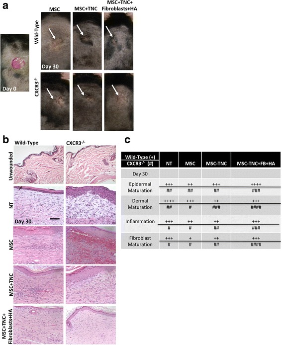
Fig. 4.

In-vivo analysis of MSC + fibroblast hydrogel matrix system. a Representative photographs of full-thickness wounds 30 days post transplantation and with MSCs, MSCs + TNC, and/or MSCs + TNC/FB with HA treatments. b H&E micrographs evaluated histologically for inflammation, and maturation of the dermis and epidermis. c Quantitative histological assessments of each wound at all time points by a blinded veterinary pathologist (day 30 shown). + Wild-type mice, # CXCR3–/–. Measurements made on a scale of 0–4 compared to unwounded within each genotype to determine scoring averages. Scale is 0–4 with +/# = 1. *p < 0.05, compared to unwounded genotype. Histological sections and data from a representative at least three mice across three experiments. Original magnifications, ×400. HA hyaluronic acid, MSC mesenchymal stem cells, NT no treatment, TNC tenascin-C
