
Fig. 5.
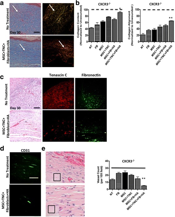
MSC + fibroblast hydrogel matrix system alters ECM remodeling outcomes. a Masson’s Trichrome staining used to detect collagen being produced, while Picrosirius Red staining used to detect appropriately aligned collagen bundles. Wounds of CXCR3–/– mice showed distinct patterns of collagen remodeling improvement when treated with MSC + TNC/fibroblast with HA treatments versus no treatment. b Wound collagen quantification of both Masson’s Trichrome and Picrosirius Red staining was done by MetaMorph imaging software. CXCR3–/– mice wounds treated with MSCs + TNC/fibroblasts with HA showed a more organized dermal matrix, longer and thicker collagen fibers, and a mature scar, as shown by arrows (determined by total integrated birefringence compared with contralateral unwounded skin) versus that of the NT. c Immunohistochemistry of matrix proteins tenascin-C and fibronectin were stained in tissue at 30 days post transplantation. d, e Angiogenesis was assessed by immunohistochemistry using CD31 marker and histologically by H&E staining. Vessels were quantified by positive stain counts. Original magnifications: a, c × 400, d × 200, H&E × 100 and × 600. *p < 0.05, **p < 0.05. Images, histological sections, and data shown from a representative at least three mice across three experiments. FB fibroblasts, HA hyaluronic acid, MSC mesenchymal stem cells, NT no treatment, TNC tenascin-C (Color figure online)