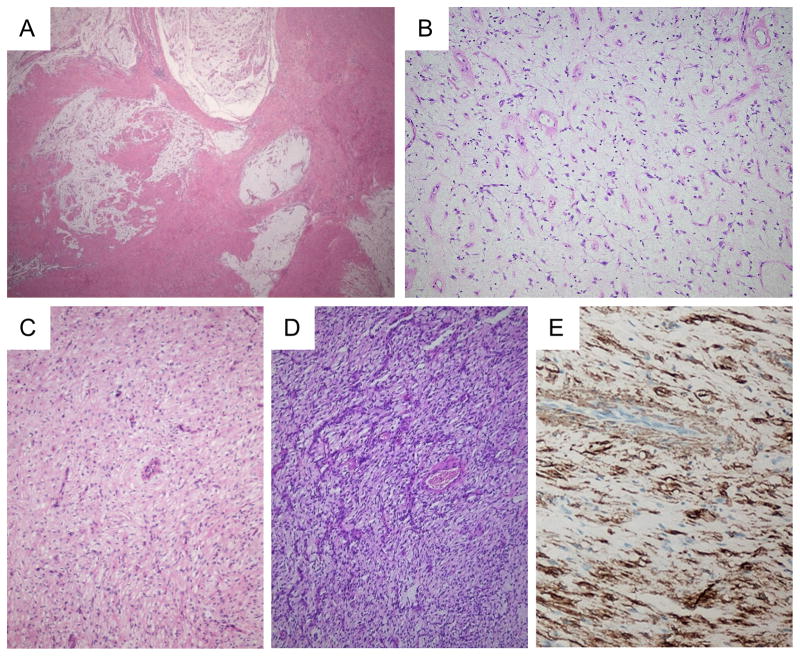
Figure 1.
Histology of plexiform fibromyxomas of the stomach. Low power view, showing the lobular and plexiform architecture (A). At high power, the myxoid matrix contains numerous vessels, bland spindle cells and scattered inflammatory cells (B). In some lesions/nodules, the cellularity is somewhat increased. Note the bland appearance of the spindle cells and the prominent vessels (C and D). Tumor cells express alpha smooth muscle actin by IHC (E).

