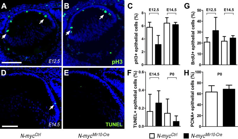
Fig. 3. N-myc-inactivation did not affect cell proliferation or cell survival in developing lens.
(A-B) Representative pictures of pH3 immunostaining in N-mycCtrl and N-mycMlr10-Cre lenses at E12.5. (C) Proportion of pH3+ epithelial cells at E12.5 (n = 3) and E14.5 (n = 3). (D-E) Representative pictures of TUNEL staining in N-mycCtrl and N-mycMlr10-Cre lenses at E14.5 (F) Proportion of TUNEL+ cells in E14.5 (n = 4) and P0 (n = 3) lenses. (G-H) Quantification of BrdU+ (n = 4) and PCNA+ (n = 8) epithelial cells in the indicated developmental stages. t-tests were performed to compare N-mycCtrl and N-mycMlr10-Cre in every graph. Error bars indicate S.D.; Scale bar: 100 µm.

