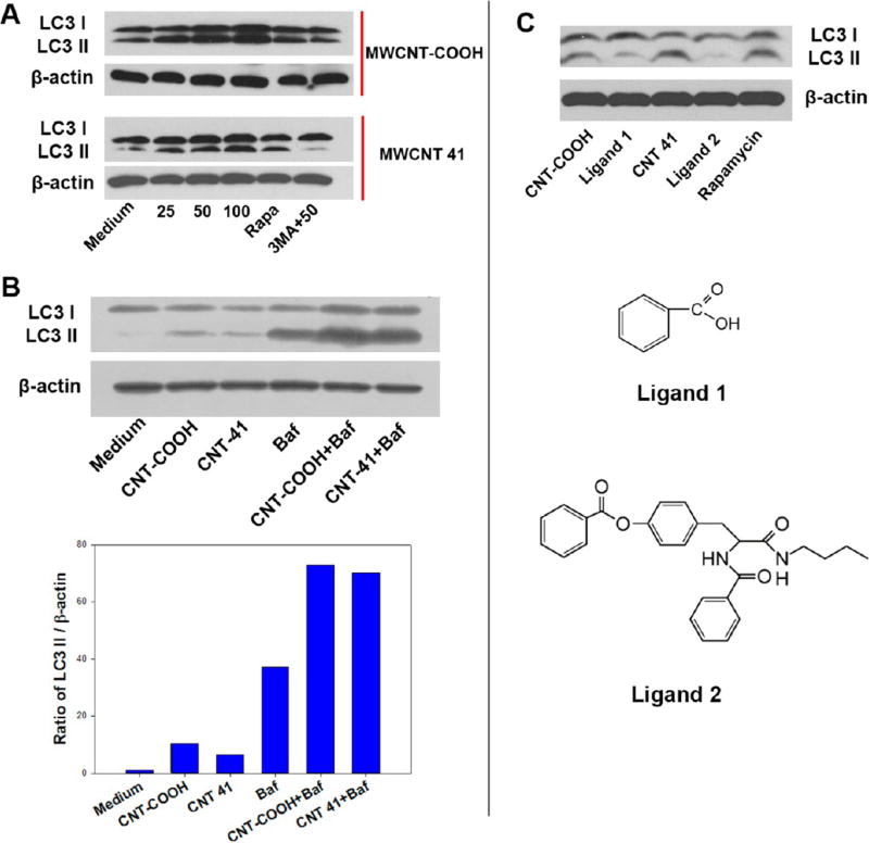
Figure 6.
Autophagy induction by MWCNTs but not ligands. In HEK 293 cells, MWCNT-COOH and MWCNT 20 induce an increased level of LC3 II isoform, while the small molecule ligands alone did not. (A) After treatment with both MWCNT-COOH and MWCNT 41 at different concentrations, the levels of LC3 I and LC3 II isoforms were detected by immunoblot assay. Rapamycin (treatment for 4 h at 4 µM) was used as the positive control. 3-Methyladenine was used as an autophagy inhibitor. (B) HEK 293 cells were treated with MWCNT-COOH or MWCNT 41 at 100 µg/mL with or without bafilomycin (100 nM) for 4 h. Cell lysate was then analyzed by Western blot against LC3B antibody. Bar chart represents the normalized band intensity ratio of LC3 II and β-actin as quantified by ImageJ. (C) Free small molecule ligands for modifying both MWCNTs surface did not cause an increase in level of LC3 II isoform using the equivalent concentration 0.04 µM. β-Actin was used as loading control.

