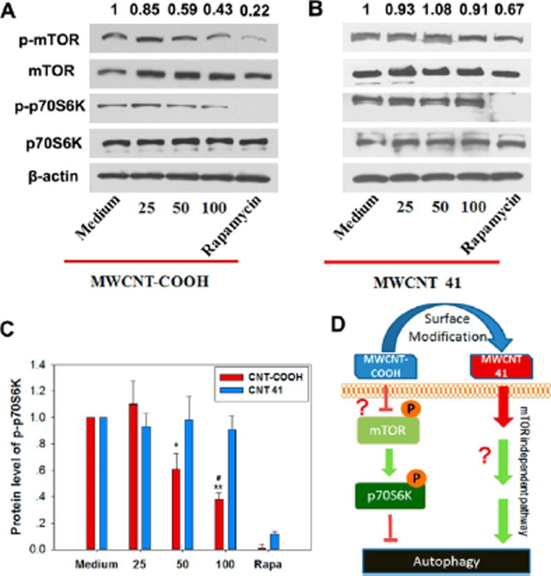
Figure 7.
mTOR-dependent and -independent autophagy induced by MWCNT-COOH and 41. HEK 293 cells were treated with MWCNTs at different concentrations for 24 h before harvesting cell lysates for immunoblot assay. Rapamycin (4 µM) was used as a positive control. β-Actin was used as a loading control. (A) The phosphorylated forms of mTOR (p-mTOR) and its substrate p70S6K (p-p70S6K) were decreased by treatment with MWCNT-COOH in a dose dependent manner. (B) In comparison, treatment with MWCNT 41 did not lead to such a change. Values above each band represent the band intensity ratio for p-mTOR/mTOR. (C) Ratio of band intensity for p-p70S6K/β-actin and p70S6K/β-actin. *P < 0.05, **P < 0.01 vs medium control, #P < 0.05 vs value at 50 µg/mL. (D) A schematic showing the action modes of MWCNT-COOH and MWCNT 41. MWCNT-COOH suppresses the phosphorylation of mTOR. This action inhibits the phosphorylation of the substrate p70S6K and therefore releases its suppression on autophagy. MWCNT 41 activates autophagy by another pathway. Question marks indicated unknown mechanism.

