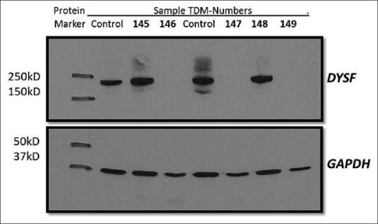
Figure 2.

Western blot showing dysferlin protein expression. Immunoblotting analysis of dysferlin protein expression. Protein lysate from control as well as patient monocytes analyzed for dysferlin expression using NCL-hamlet antibody specific for 235 kDa dysferlin protein. Lane 2 and 5 control lysate shows an intense band. Lane 4, 6 and 8 shows absence of dysferlin protein in TDM-146 (JF tool results 97.34% probability with high concordance), TDM-147 (JF tool results 97.28% probability with high concordance), TDM-149 (JF tool results 64.9% probability with medium concordance) respectively. Lane 3 and 7 shows presence of dysferlin protein in TDM-145 (JF tool result 52.2% probability with low concordance) and TDM-148 (JF tool results 95.11% probability with high concordance). GAPDH used as a loading control (37 kDa)
