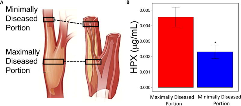
Figure 4. Hemopexin content is higher in maximally diseased carotid artery plaque segments.
(A) Anatomical distinction between maximally and minimally diseased plaque segments at the extracranial carotid artery bifurcation. (B) Difference in hemopexin content between maximally and minimally diseased carotid artery segments. * p < 0.05

