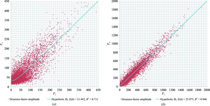
Figure 6.
F o versus F c graphs for (a) electron diffraction of a single lysozyme nanocrystal and (b) an X-ray data set for cubic (bovine) insulin at 1.6 Å resolution. The data were least-squares fitted with a hyperbolic function described by 〈|F o|〉 = [|F c|2 + 〈|E(h)|〉2]1/2. F o versus F c graphs for only the low-resolution part of the single-crystal data and for the merged crystal data are shown in Supplementary Fig. S6.

