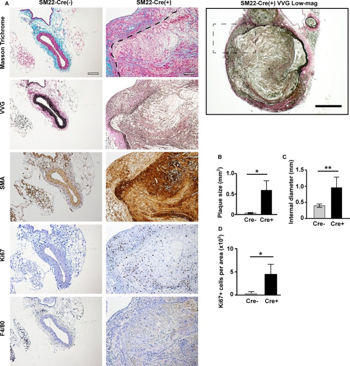
Figure 4.

The loss of vascular wall integrity and vascular smooth muscle cell (VSMC) proliferation in VSMC MT1‐MMP (membrane‐type 1 matrix metalloproteinase) knockout mice. A, Atherosclerotic aneurysms found in iliac arteries of SM22α‐Cre(+)MMP14 F/ FA poe −/− mice. Masson's Trichrome staining; the border between the vascular wall and the atheroma is demarcated with a dashed line. Verhoeff–Van Gieson staining (VVG), smooth muscle actin (SMA) staining, Ki67 staining, and F4/80 (a macrophage marker) staining. Scale=100 μm. The lower magnification (×4) micrograph of SM22α‐Cre(+)MMP14 F/ FA poe −/− VVG staining on the right. Scale=200 μm. The lesion in the dashed square is shown on the left. B, Plaque area. C, The internal diameter of iliac arteries. D, Ki67‐positive area. Mean±SEM; n=5 and n=7 for each group. One‐way ANOVA. *P<0.05, **P<0.005.
