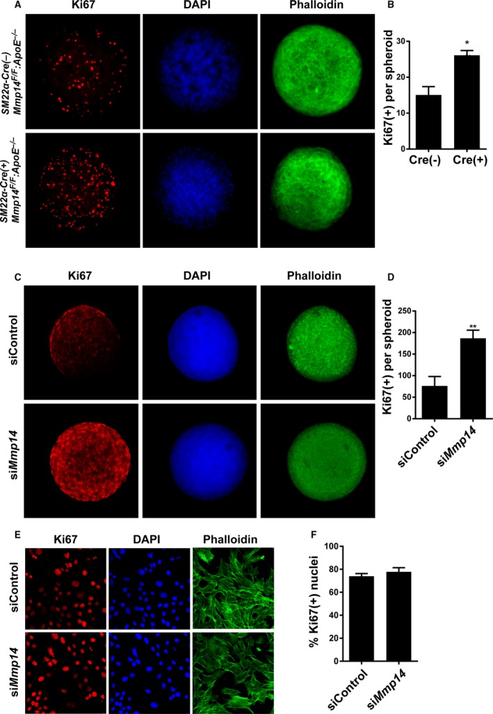
Figure 6.

MT1‐MMP (membrane‐type 1 matrix metalloproteinase) limits vascular smooth muscle cell (VSMC) proliferation in 3‐dimensional (3‐D) organoids. A, Primary VSMCs isolated from Cre(−)Mmp14 F/ FA poe −/− mice and Cre(+)Mmp14 F/ FA poe −/− mice were cultured as a 3‐D spheroids for 48 hours and stained for Ki67 (red), nuclei (DAPI, blue), and actin (phalloidin, green). B, Quantified intensity of Ki67 staining per spheroid. n=5 to 7. *P<0.05. C, Immortalized mouse VSMCs (MOVAS) transiently transfected with small interfering RNA (siRNA) control (siControl) and MT1‐MMP siRNA (siMmp14). Ki67 (red), nuclei (blue), actin (green). D, Ki67 staining intensity per spheroid of MOVAS. **P<0.005. E, 2‐D cultured MOVAS transfected with control and MT1‐MMP siRNA. Ki67 (red), nucleus (blue), and actin (green). F, Ki67‐positive nuclei per total nuclei count.
