Figure 4.
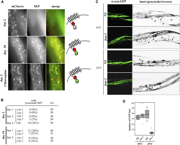
Protein aggregation in muscle of postreproductive worms. (A) Alkalinization of lysosomes in muscle cells of day 2 and day 10 wild-type animals expressing SEP::mCherry::LAAT-1. SEP fluorescence is quenched in acidic environments but detectable at pHs >6.5 (diagram on the right). Treatment with 200 mM of chloroquine leads to SEP-positive lysosomes in day 2. (B) Quantification of animals with lysosomal SEP signal in muscle cells. Two experiments (exp) with each of two independently generated transgenic lines (lines 1 and 2) were performed. (C) The in situ steady state of α-syn::GFP aggregates in muscle cells of wild-type (N2) and reproductively active (16°) or feminized (25°) fem-2(b245ts) day 6 worms. Insets are shown as grayscale/inverted images to highlight α-synuclein foci in black, denoted by arrowheads. (D) Quantification of α-syn::GFP foci (n = 20).

