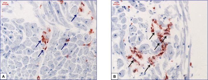
Figure 3.

Representative images of immunohistological staining from frozen samples. A and B, Increased perforin‐positive cardiac cell infiltration with focal infiltration pattern and partially beginning of cardiomyocyte destruction in a patient with perforin‐positive inflammatory cardiomyopathy (×400).
