Figure 3.

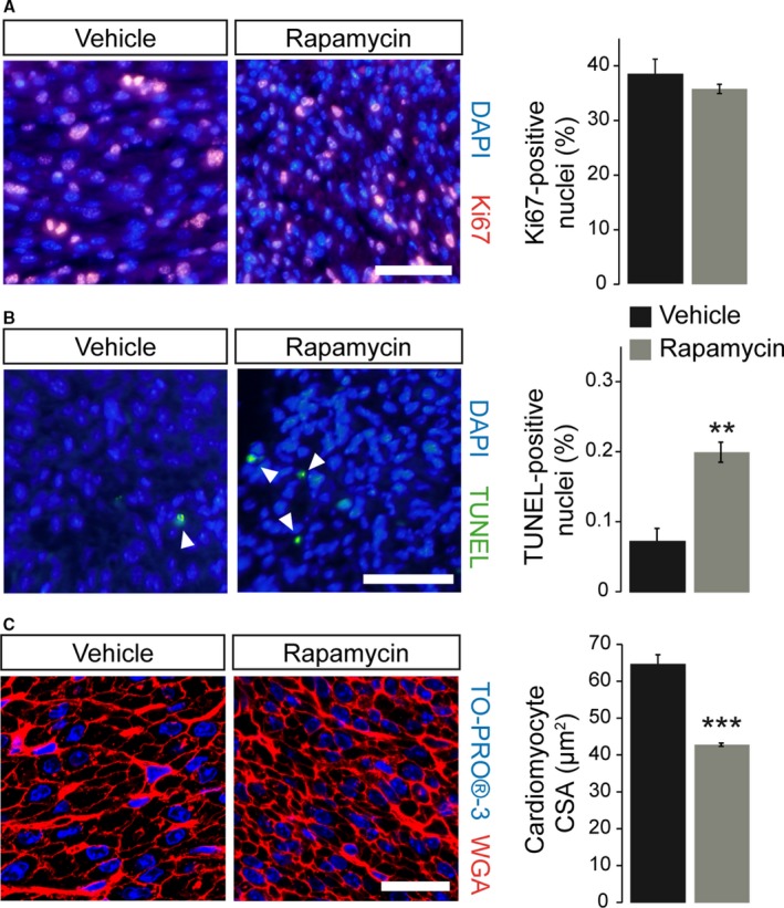
Prenatal rapamycin treatment reduces cardiomyocyte size and induces apoptosis but does not affect proliferation in the postnatal day‐1 heart. A, Quantification of immunofluorescence images of Ki67‐labeled nuclei (red) revealed unchanged proliferation rates within the left ventricular (LV) myocardium of vehicle‐ and rapamycin‐treated neonatal hearts. Nuclei were stained in blue with DAPI (scale bar=50 μm, vehicle n=6, rapamycin n=12). B, Quantification of TUNEL‐positive nuclei (green, see arrowheads) revealed significantly increased apoptosis within the LV myocardium of neonatal hearts after prenatal mTORC1 inhibition compared to vehicle‐treated controls. Nuclei were stained in blue with DAPI (scale bar=50 μm, vehicle n=4, rapamycin n=6). C, Fluorescence staining of cardiomyocyte membranes with wheat germ agglutinin (WGA, red) within the LV myocardium revealed a significantly reduced cardiomyocyte cross‐sectional area (CSA) in rapamycin‐ compared to vehicle‐treated neonates. Nuclei were stained in blue with TO‐PRO‐3 (confocal microscopy, scale bar=25 μm, vehicle n=5, rapamycin n=3). (***P<0.001, **P<0.01). TUNEL indicates terminal deoxynucleotidyl transferase dUTP nick end labeling.
