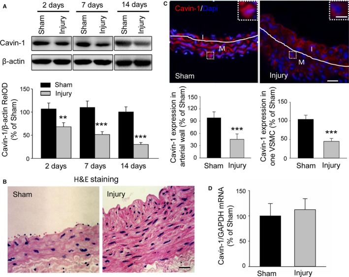
Figure 1.

Balloon injury results in a decline of Cavin‐1 protein expression but not mRNA in injured carotid arteries. A, Western blot showing the effect of balloon injury on Cavin‐1 protein expression at 2, 7, and 14 days after balloon injury or sham operation to injured carotid artery. Cavin‐1 relative optical density (RelOD) indicates the percentage of the relative optical density value of Cavin‐1/β‐actin to sham group. n=3 to 4 mice/per group, **P<0.01, ***P<0.001, vs sham group. B, Typical photographs of hematoxylin and eosin (H&E) staining showing the vascular structure of carotid at 14 days in sham and injured groups. Scale bar: 25 μm. C, Representative immunofluorescent staining and the statistical histogram showing the expression of Cavin‐1 in the injured carotid arterial wall or in 1 single vascular smooth muscle cell (VSMC) (inset) at 14 days after sham operation or left balloon injury. Red: Cavin‐1, blue: 4',6‐ diamidino‐2‐phenylindole (Dapi). The white lines show the luminal border between the intima (I) or media (M) layer. The insets at the top right corner show higher‐magnification from the white boxes in the large images. Scale bars: 25 μm, inset: 5 μm. ***P<0.001, vs sham group, n=3 to 4 mice/per group and 3 to 5 images/per mice, and n=7 to 8 cells/per image. D, Cavin‐1 mRNA in carotid artery from sham or balloon‐injured group was determined by real‐time reverse transcriptase polymerase chain reaction (n=6–8).
