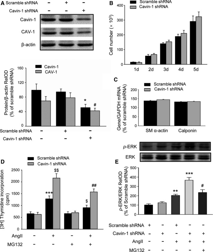
Figure 4.

Inhibition of Cavin‐1 expression by shRNA promotes AngII‐induced VSMC proliferation via further activation of extracellular signal‐regulated kinase (ERK). A, Representative Western blot and statistical analysis showing the efficiency of Cavin‐1 shRNA on the inhibition of Cavin‐1 expression and even on CAV‐1 expression in VSMC culture (n=3–4/per group, Cavin‐1 expression: *P<0.05 vs Scramble shRNA alone; CAV‐1 expression: # P<0.05 vs Scramble shRNA alone). B, The histogram of VSMC number showing the effect of Cavin‐1 shRNA on VSMCs proliferation in normal growth culture medium (10% FBS DMEM). C, Knockdown of Cavin‐1 protein expression by Cavin‐1 shRNA had no effect on VSMC phenotypic marker α‐SM actin and calponin (n=6–8/per group). D, [3H]‐thymidine incorporation measured as count per minute (cpm) showing the effect of Cavin‐1 knockdown on VSMC proliferation in stimulation with AngII (100 nmol/L) for 24 hours with or without pretreatment with proteasome inhibitor MG132 (10 μmol/L) for 1 hour. n=9, ***P<0.001 vs Scramble shRNA alone, $ P<0.05, $$ P<0.01 vs Scramble shRNA treated with AngII and ## P<0.01 vs Cavin‐1 shRNA treat with AngII. E, Western blot showing the effect of Cavin‐1 knockdown on AngII‐induced ERK phosphorylation (p‐ERK) with or without pretreatment with MG132. p‐ERK/ERK RelOD indicated the percentage of the relative optical density value of p‐ERK/ERK to Scramble shRNA group. n=3, **P<0.01, ***P<0.001 vs Scramble shRNA treated with AngII, # P<0.05 vs Cavin‐1 shRNA treated with AngII. Ang II indicates angiotensin II; FBS, fetal bovine serum; RelOD, relative optical density; shRNA, short hairpin RNA; VSMCs, vascular smooth muscle cells.
