Figure 6.

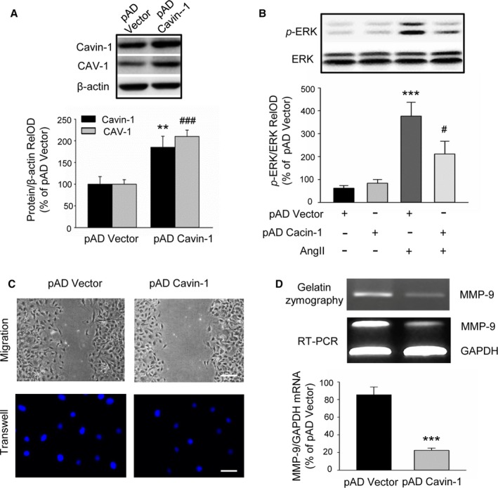
Effect of Cavin‐1 overexpression on VSMC proliferation and migration. A, Representative Western blot and statistical analysis showing the efficiency of adenovirus‐mediated Cavin‐1 cDNA on Cavin‐1 expression and even on CAV‐1 expression (n=3, **P<0.01 vs pAD‐Vector, Cavin‐1; ### P<0.001 vs pAD‐Vector,CAV‐1). B, p‐ERK were markedly increased after treatment VSMC with AngII (100 nmol/L) for 30 minutes, but significantly reduced by Cavin‐1 overexpression. Only overexpression of Cavin‐1 had no effect on VSMC ERK activity (n=3, ***P<0.001 vs pAD alone, # P<0.05 vs pAD Vector plus AngII). C, The scratch assay and transwell experiment showing the effect of overexpression Cavin‐1 on VSMC migration. Scale bars: upper, 50 μm, lower, 25 μm. D, The results from gelatin zymography and RT‐PCR showing the change of MMP‐9 protein and mRNA after Cavin‐1 overexpression. The 48‐hour conditioned medium from VSMCs transiently transfected with pAD Vector or pAD Cavin‐1 was collected and equal amounts of protein were subjected to gelatin zymography. Equal amounts of mRNA from VSMCs transfected with pAD Vector or pAD Cavin‐1 were subjected to RT‐PCR using primers to identify MMP‐9 or GAPDH. PCR products of the expected size were identified by agarose gel electrophoresis (n=6, ***P<0.001 vs pAD Vector). Ang II indicates angiotensin II; MMP‐9, matrix‐degrading metalloproteinase‐9; pAD, adenoviral plasmid; p‐ERK, extracellular signal‐related kinase phosphorylation; RelOD, relative optical density; RT‐PCR, reverse transcriptase polymerase chain reaction; VSMCs, vascular smooth muscle cells.
