
Figure 7.
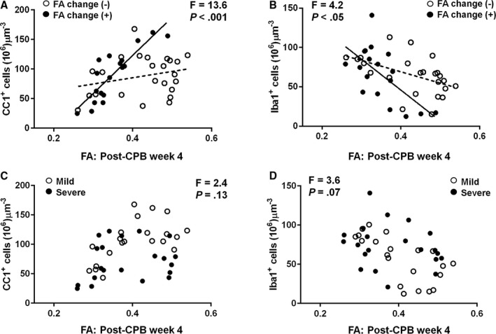
FA is a powerful biomarker to define WM cellular alterations after cardiac surgery. A and B, When the correlation is compared in WM areas between with and without FA changes (BCC, SFWM, MFWM vs ALIC, M‐PVWM, L‐PVWM, IFWM), better fits in positive FA relation with mature oligodendrocytes (A) and negative association with microglia (B) are observed in the area with FA change (black line) compared with the WM without change (dotted line), suggesting that FA changes on postoperative week 4 enable identification of CPB‐induced cellular alterations (n=6 animals). C and D, There is an unchanged relation between FA and the number of mature oligodendrocyte cells (C) and microglia (D) between mild and severe CPB groups (n=6 animals). ALIC indicates anterior limb of the internal capsule; BCC, body of the corpus callosum; CPB, cardiopulmonary bypass; FA, fractional anisotropy; IFWM, inferior frontal white matter; L‐PVWM, lateral periventricular white matter; MFWM, middle frontal white matter; M‐PVWM, medial periventricular white matter; SFWM, superior frontal white matter; WM, white matter.