Figure 4.

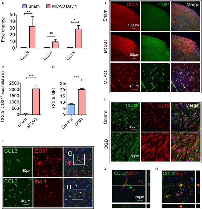
CCR5 ligands are upregulated in the ischemic brain. Cerebral ischemia was induced by 60 minutes of transient middle cerebral artery occlusion MCAO. Animals were euthanized 1 day after MCAO or sham operation. A, The mRNA expression of CCL3, CCL4, and CCL5 was measured by real‐time polymerase chain reaction. B, Representative double immunofluorescence staining of CCL5 and CD31. Images are representative of 3 to 5 animals/group. C, Quantification of CCL5+ CD31+ blood vessel length in sham and tMCAO brains, n=3 to 5/group. D and E, Cultured endothelial cells were challenged with oxygen‐glucose deprivation (OGD) for 4 hours. The cells were fixed and stained for CCL5 and CD31. D, Quantification of mean fluorescence intensity (MFI) of CCL5 in the control or OGD‐challenged endothelial cells. E, Representative CCL5 and CD31 immunofluorescent staining images of control or OGD‐challenged endothelial cells. F, Representative images of CCL3/CD31 and CCL3/Iba‐1 double staining. The white boxes are enlarged (G and H). Images are representative of 3 to 5 animals/group. G, High‐power confocal z‐stack image of CCL3/CD31 double staining, n=5/group. H, High‐power confocal z‐stack image of CCL3/Iba‐1 double staining. *P≤0.05, **P≤0.01, ***P≤0.001. CCL indicates ligand for CCR; CCR5, C‐C chemokine receptor type 5; Treg, regulatory T cell.
