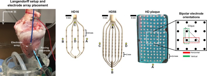
Figure 1.

Langendorff setup and 2‐dimensional electrode arrays: HD16, HD56, and HD plaque arrays. An experimental setup is shown to establish axes relative to the Langendorff suspension as well as the placement of electrode arrays and pacing electrodes. Two‐dimensional electrode arrays laid and stitched flat onto a ventricular surface were used for electrical mapping. Stars indicate the pacing sites chosen for each electrode array. HD16 was used for rabbit hearts, HD56 for porcine hearts, and HD plaque for human hearts. By subtraction, bipolar voltage maps were created for 2 orthogonal electrode orientations (horizontal and vertical). Omnipolar electrograms were derived from a group of 4 adjacent electrodes in a triangular configuration (clique) of each electrode array.
