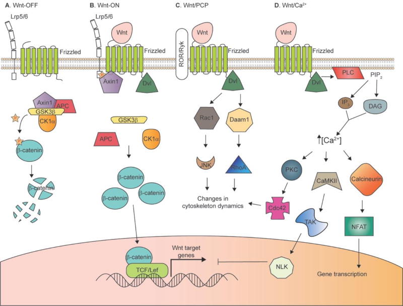
Figure 1. Wnt signaling pathways.
A and B: Canonical (β-catenin-dependent) pathway. In the “Wnt-OFF” state (A), β-catenin levels are kept low in the cytoplasm by the action of the “destruction complex”. GSK3β phosphorylates β-catenin, which targets it for desctruction by the proteasome. Wnt binding to Frizzled and Lrp5/6 co-receptors results in disruption of the destruction complex, allowing β-catenin to accumulate in the cytoplasm. β-catenin can then translocate to the nucleus and promote the activation of Wnt target genes by displacing co-repressors, and recruiting co-activators, of TCF/Lef. C: Wnt/PCP pathway. Binding of Wnt to Frizzled and ROR or Ryk co-receptors leads to recruitment of Dvl, which can act through Rac1 or Daam1 to initate changes in cytoskeletal dynamics important for cell orientation and movement. D: Wnt/Ca2+ pathway. Wnt binding and recruitment of Dvl leads to activation of PLC, which cleaves PIP2 to generate IP3 and DAG. This leads to release of Ca2+ from intracellular stores, and downstream signaling through a number of Ca2+-dependent kinases and phosphatases.

