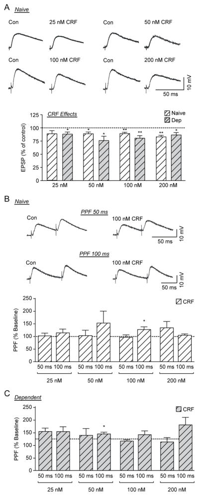
Figure 2. CRF decreases evoked EPSP amplitude in CeA neurons of naïve and ethanol-dependent rats.

A) Top: Representative recordings of evoked EPSPs in CeA neurons from naïve rats before and during CRF application. Bottom: Histograms representing percent peak decrease in evoked (at half max stimulus intensity) EPSP amplitudes during superfusion of different concentrations (25, 50, 100 and 200 nM) of CRF on the CeA of naïve and ethanol-dependent rats (6–16 cells from at least 4 naïve or dependent rats per group). B) Top: Representative recordings of PPF at both 50 ms (top traces) and 100 ms (bottom traces) ISI in a CeA neuron from a naïve rat before and during superfusion of 100 nM CRF. Bottom: Histograms representing PPF ratio as a percentage of baseline after superfusion of 4 concentrations of CRF at 50 ms and 100 ms ISI in naïve rats. CRF significantly increased the PPF ratio of evoked EPSPs at the 100 ms ISI with 100 nM CRF C) Histograms representing PPF ratio as a percentage of baseline after superfusion of 4 concentrations of CRF at 50 ms and 100 ms ISI in ethanol-dependent rats. CRF significantly increased the PPF ratio of evoked EPSPs at the 100 ms ISI with 50 nM CRF. *p <0.05 and **p<0.01 by one-sample t-test.
