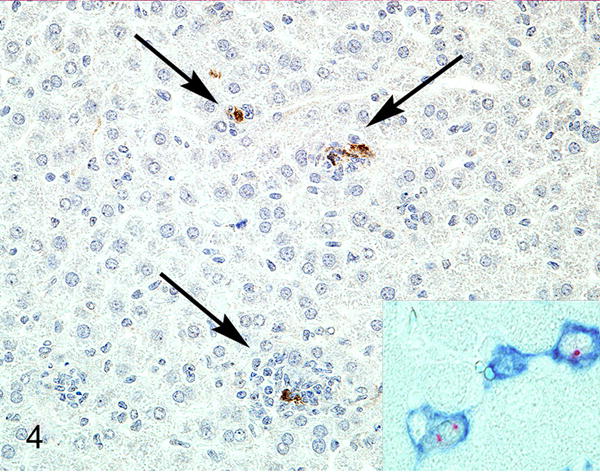
Figure 4.

Liver, 2.5-month-old Rag1−/−/IFNγR−/− mouse. There is expression of MNV-1 ProPol in inflammatory cells (arrows). Inset: hepatitis, liver, Rag1−/−/STAT1−/− mouse. The sinusoidal lining cells have F4/80-positive cell membranes (blue) and MNV ProPol labeling (red) of the cytoplasm. Immunohistochemistry for MNV-1 ProPol. Figures 3 and 4 are reprinted from “Pathology of immunodeficient mice with naturally occurring murine norovirus infection,” Ward JM, et al. Toxicol Pathol. 2006;34(6): 708–15 with permission from SAGE Publications.
