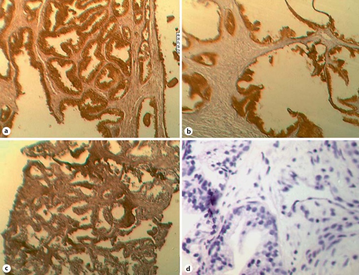
Fig. 1.
Photomicrographs of immunoreactivity of Aurora kinases and frequency. a AURKA-positive BPH (dark-brown stain), ×400 magnification. Epithelial cells are predominantly stained. The IHC score was 5+. b AURKB-positive BPH (dark-brown stain), ×400 magnification. The IHC score was 4+. c AURKC-positive PCa (dark-brown stain), ×400 magnification. The IHC score was 3+. d Negative control slide (blue nucleic stain), ×400 magnification. The IHC score was 0. Scale: 1 cm = 18 µM in ×400 magnification.

