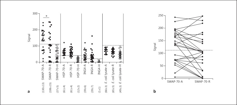
Fig. 1.
a ELISA detection of IgG antibodies (fluorescence signal values) to SWAP-70, HSP-70, ING4 and the E. coli lysate in the sera of healthy controls (H) and relapsing RRMS patients during attack (A) and remission (R) periods. Horizontal lines indicate the mean values of each group, and the numbers in parentheses indicate the average ± SE for each group. * p < 0.05. b Comparison of serum SWAP-70 antibody levels of RRMS patients during attack (A) and remission (R) periods. The dashed lines represent the cut-off value for SWAP-70 antibody seropositivity (2 SD above the mean of healthy controls).

