Fig. 2.

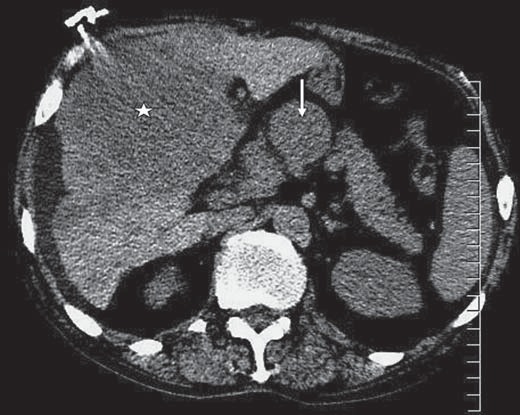
Axial noncontrast CT image through the upper abdomen shows a large heterogeneous mass of relatively low attenuation within a large portion of the liver (white star) and portahepatis lymphadenopathy (white arrow).

Axial noncontrast CT image through the upper abdomen shows a large heterogeneous mass of relatively low attenuation within a large portion of the liver (white star) and portahepatis lymphadenopathy (white arrow).