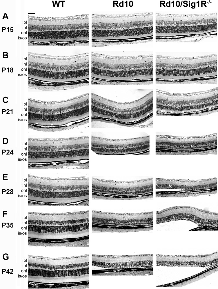
Figure 4.
Retinal structure and morphometric analysis. Retinal sections of eyes embedded in JB4 and stained with H&E from WT, rd10, and rd10/Sig1R−/− mice. (A–G) Representative image from WT, rd10, rd10/Sig1R−/− groups at P15 (A), P18 (B), P21 (C), P24 (D), P28 (E), P35 (F) and P42 (G). Note accelerated retinal detachment and paucity of PRC in the ONL in rd10/Sig1R−/− compared with rd10 mice. (H–Y) Morphometric analyses of TRT (H–M), ONL thickness (N–S), and OPL thickness (T–Y) at P18, P21, P24, P28, P35, and P42 separately. gcl, ganglion cell layer; ipl, inner plexiform layer; inl, inner nuclear layer; opl, outer plexiform layer; onl, outer nuclear layer; is, inner segment; os, outer segment; rpe, retinal pigment epithelium. Data are the mean ± SEM of measurements from six to nine mice per group. *P < 0.05; **P < 0.01; ***P < 0.001. Scale bar: 50 μm. Numbers of mice used in the analysis are provided in Supplementary Table S1.

