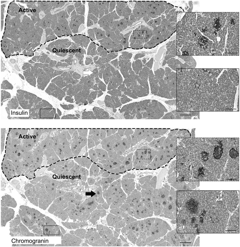
Figure 1.
The mosaic arrangement of the endocrine pancreas in CHI-A. Within the dotted region of the pancreas in the upper panel (Active), islets rich with insulin-positive staining are clearly visible. The equivalent islet structures are also visible in the serial section of tissue shown in the lower panel, which was stained for chromogranin A. However, in the area outside the dotted region, insulin expression is weak, suggesting that islets within this domain are limited in β-cell numbers and/or are quiescent (Quiescent). Within the quiescent domain, islets are clearly present when stained for the neuroendocrine marker chromogranin A. The arrow and boxed regions show how islets are clearly visible when stained for chromogranin but not insulin. Scale bar = 500 μm; boxed regions = 50 μm.

