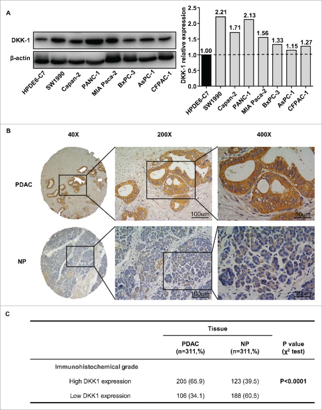
Figure 2.

DKK-1 expression in PDAC tissue at protein level. (A) Western blot of 8 PDAC cell lines and 1 nonmalignant HPDE6-C7 cells and the quantification of western blot demonstrated that expression of DKK-1 was increased in PDAC cell lines. (B) Typical imagine of DKK-1 expression in PDAC tissue microarray by IHC. (C) In Ren Ji cohort 2, the rate of high DKK-1 expression was significantly higher in PDAC group compared with NP group.
