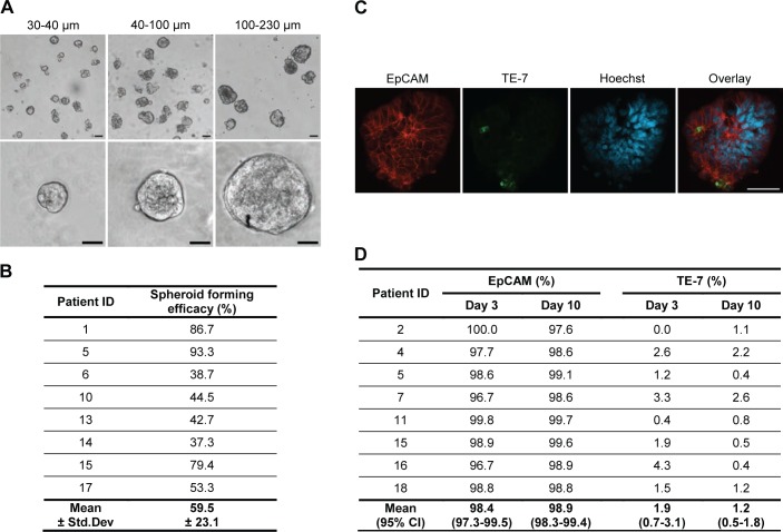
Fig 1. Colorectal spheroid cultures predominantly consist of epithelial cells.
(A) Spheroids of three different sizes at low and high magnification after 3 days of culture. Size bar = 50 μm. (B) Spheroid forming efficacy of isolated tumour fragments. Spheroid forming efficacy was defined as the percentage of isolated tumour fragments that had formed spheroids within 3 days of culture. (C) Immunostaining of spheroids for epithelial cell marker EpCAM (red) and fibroblast marker TE-7 (green) after 10 days of culture. Nuclei are stained with Hoechst (blue). Size bars = 50 μm. (D) No significant difference in percentage of spheroid cells stained for EpCAM (p = 0.387) and TE-7 (p = 0.196) at day 3 and day 10 was observed.

