
Fig. 2. VSMCs align weakly on cylinders when isolated.
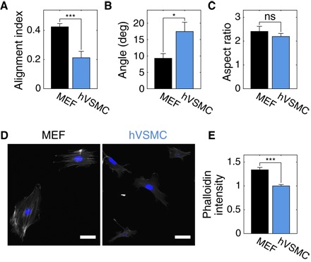
(A) Isolated hVSMCs have a smaller AI than isolated MEFs on cylinders with Rc = 40 μm; this is due to weaker axial alignment (B) and not a difference in elongation (C), as measured from the dimensions of ellipses fit to cell outlines. At least 16 cells were analyzed in each condition. Representative images of phalloidin-TRITC (gray) and 4′,6-diamidino-2-phenylindole (DAPI; blue) staining of MEFs and hVSMCs on coverslips (D) and corresponding phalloidin-TRITC intensities (E). Scale bars, 50 μm. At least 40 cells of each type in two independent experiments were analyzed. Results are mean and SE. *P < 0.05, ***P < 0.001, Student’s t test.